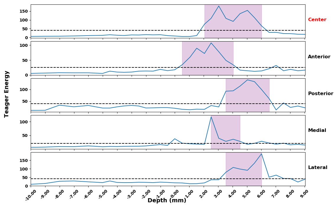
The figure above shows an example feature, Teager energy, extracted from the raw brain recording data. The depth values indicate distance from the surgically planned target (negative values above, positive values below), there are 5 electrodes used during the recording. This figure illustrates that as the electrodes enter the STN, the Teager energy increases above the avergae (dotted line) and indidates that the electrodes have entered a nucleus. Using various features, extracted from the raw signal, this project aims to build a predictive model that informs the surgeon of the most optimal surgical implantation site.
The ability to train a predictive model that is capable of estimating the dorsal/ventral borders of the STN, as well as the sensorimotor aspect, would reduce surgery time and improve patient outcome.